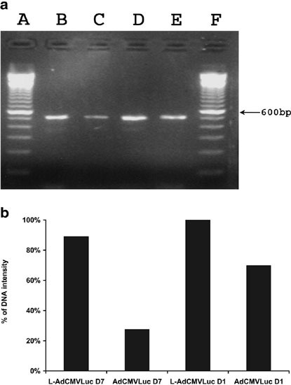
Figure 7.

PCR analysis of the amount of viral DNA in lungs at days 1 and 7 following tail vein injection of AdCMVLuc alone or DOTAP:cholesterol liposomes followed by AdCMVLuc. Mice received tail vein injection of AdCMVLuc, alone or following the injection of DOTAP:cholesterol liposomes. At days 1 and 7, mice were killed and lungs were collected. Genomic DNA was extracted from the lungs and used as the template. Luciferase cDNA-specific primers used in the PCR amplification were LUC-1 (5′-CGTCACATCTCATCTACCTC-3′) and LUC-2 (5′-GTATCCCTGGAAGATGGAAG-3’), which generated a 510-bp fragment as shown in (a). (a and f) 100 bp DNA marker; (b) L-AdCMVLuc (day 7); (c) AdCMVLuc (day 7); (d) L-AdCMVLuc (day 1); (e) AdCMVLuc (day 1). The results of densitometry analysis of the amplified fragments were shown in (b) and were expressed as the percentage of the intensity of the DNA fragment on lane D.
