FIGURE 2.
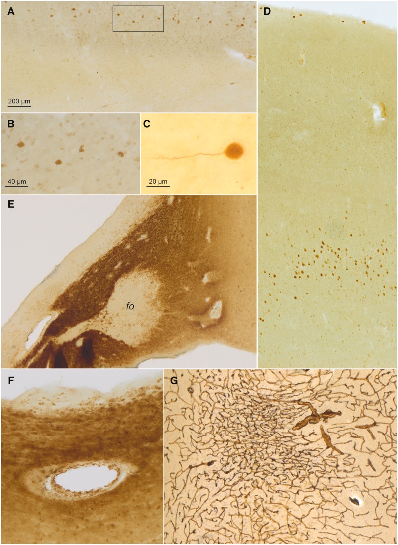
CD77-immunopositive intraneuronal inclusions (continued), CD77-immunopositive astrocytic and vascular inclusions in a 58-year-old male with Fabry disease. (A) Cajal-Retzius cells in layer I of the anterior cingulate gyrus contained Gb3 accumulations (DAB, brown chromogen). Framed area is shown at higher magnification in panel (B). (C) Detail of SMI-311-immunopositive Cajal-Retzius cell in the molecular layer located between the dentate fascia and sector CA1 close to the obliterated hippocampal fissure. (D) Overview showing CD77-positive (DAB) pyramidal cells in layers I and Vb of the fusiform gyrus. (E) Intensely CD77-immunoreactive astrocytes in the hypothalamus and, to a considerably lesser extent, in the fornix (fo). (F) Gb3 (CD77) deposits (DAB, brown chromogen) in a vessel wall close to the lateral ventricle surrounded by intensely CD77-positive astrocytes. (G) Tortuous vessels and an infarction marked by puckering of the capillary network (collagen IV) in the subiculum (DAB, brown chromogen, plus erythrosin-phosphotungstic acid-aniline blue staining) (96). Immunoreactions: CD77 (DAB) or, in (G), collagen IV with EPA staining in 100 µm sections. Scale bar in (A) applies to (D), (E), and (G). Scale bar in (B) is also valid for (F).

