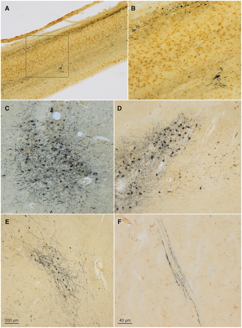
FIGURE 3.
α-Synuclein-immunopositive Lewy neurites and Lewy bodies in a 58-year-old male with Fabry disease. (A) In the olfactory bulb, syn-1 immunoreactive LP (SK-4700, dark blue chromogen) occurred in the olfactory tract and anterior olfactory nucleus, and numerous CD77-positive (DAB, brown chromogen) astrocytes were present. Framed area is shown at higher magnification in panel (B). (C) Micrograph of LP and cell loss in the locus coeruleus. (D) LP (SK-4700) accompanied by very severe cell loss in the pars compacta of the substantia nigra (at left); noncatecholaminergic nerve cells within the paranigral nucleus (at right) were CD77-immunopositive (DAB) and accompanied by fewer LP-containing catecholaminergic neurons than the pars compacta. (E) Lewy bodies/neurites (SK-4700) in the somatomotor system tegmental pedunculopontine nucleus. (F) LP (SK-4700) in axons of the vagal nerve in the medulla oblongata (intermediate reticular zone), a finding identical to that seen in the medulla of individuals with Parkinson’s disease. Immunoreactions: CD77 or CD77 plus syn-1 in 100 µm sections. Scale bar in (E) applies to (A), (C), and (D). Scale bar in (F) is also valid for (B).

