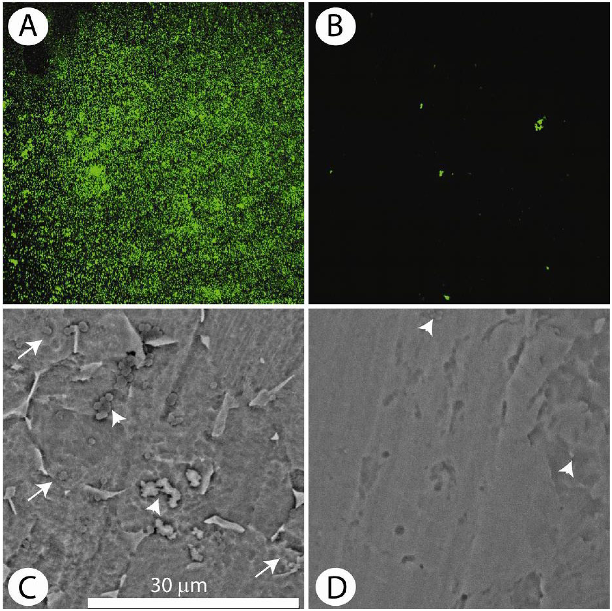
Fig. 2.

S. aureus accumulation on the vancomycin (VAN-Ti) tethered surface. (A) Distribution of bacteria (103 cfu/mL, 24 h) stained with the Live/Dead assay on the control surface (no antibiotic) and visualised by confocal microscopy. The high green fluorescence indicates extensive bacterial colonisation. (B) Distribution of bacteria on the tethered antibiotic surface (VAN-Ti) stained with the Live/Dead assay. Note the absence of green fluorescence indicating minimal adherent bacteria. (C) and (D) SEM of control and treated titanium surfaces. (C) SEM of the control surface following incubation with bacteria for 24 h. Arrowheads show the presence of small colonies of bacteria; arrows indicate biofilm encased bacteria. (D) Vancomycin treated titanium surface. Note the few bacteria on the treated
