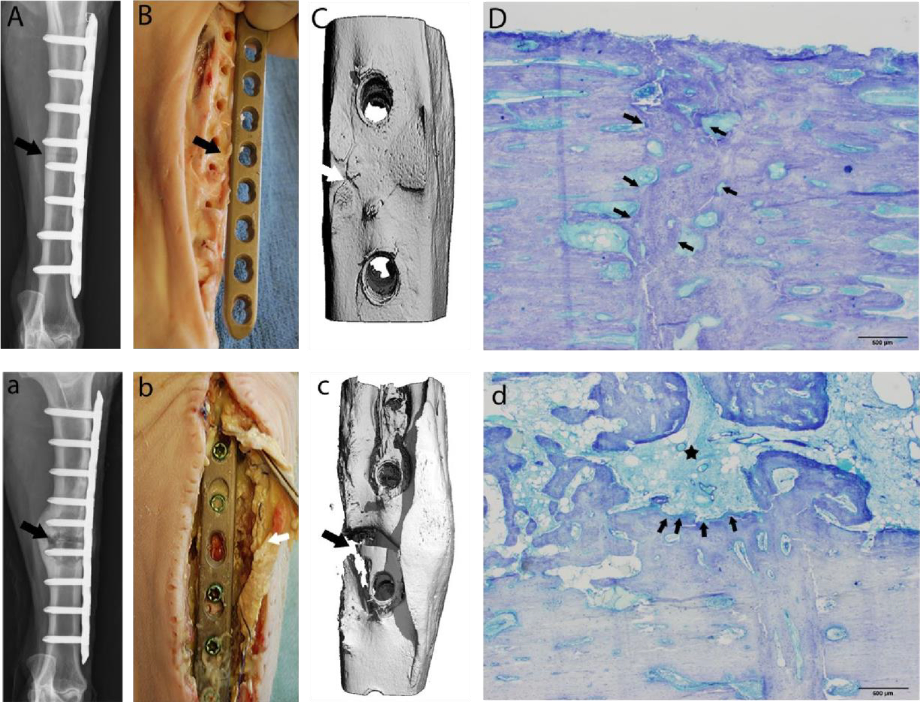
Fig. 4.

Representative images at 90 days postoperative. (A-D) Tissue repaired with VAN-LCP (vancomycin-locking compression plate); (a-d): repair conducted with unaltered LCP): Digital radiographs with cranio-caudal view of osteotomy site confirming persistence of the osteotomy (a: arrow), and a healing osteotomy site (A: arrow). 3D reconstructed micro-CT views (Bb) of the osteotomy site correlating with digital radiographs Aa. Note the persistence of the osteotomy gap (arrows: a & b). There was evidence of abscessation and an unstable osteotomy (c: arrow) during aseptic tissue harvest in the control cohort animals, while the tibiae repaired with coated plates presented with a grossly stable fracture callus and no evidence of infection (C: arrow). The histology images (Dd) corroborate the gross anatomic and radiographic findings. A representative toluidine blue-stained, mid-sagittal section from the treatment cohort. Scale bar = 500 μm. (D) comprising of the near cortex of the osteotomy site (row of arrows) is completely bridged by remodelling bone with minimal endosteal and periosteal callus formation. A photomicrograph (d) of a toluidine blue-stained, mid-sagittal section comprising the near cortex from sheep receiving an unaltered cpTi LCP demonstrates severe cortical lysis (arrows) with central cavity containing debris and pockets of suppurative inflammation with formation of large, poorly organised endosteal callus (asterisk). Scale bar = 500 μm.
