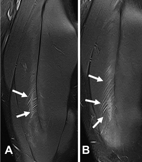
Figure 4.

A case of intramuscular injury in the rectus femoris (arrows). (A) Coronal and (B) sagittal T2-weighted fat-saturated magnetic resonance imaging scans. This type of injury entails no direct or indirect involvement of the tendon or aponeurosis, even though the injury has actually been caused by the traction of a myoconnective junction, in this case by the central tendon of the rectus femoris.
