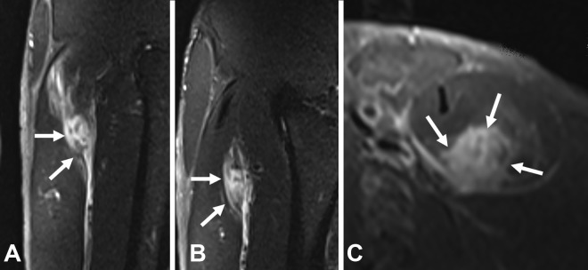
Figure 5.
A case of myofascial injury close to the proximal part of the posterior aponeurosis of the rectus femoris (arrows). (A) Coronal, (B) sagittal, and (C) axial T2-weighted fat-saturated magnetic resonance imaging scans. In this case, since the epimysium is intact, bleeding occurs in the muscle but is limited by the epimysial fascia.

