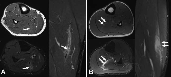
Figure 7.
Two different cases of central tendon injury of the soleus. (A) Axial T1-weighted and axial and sagittal T2-weighted fat-saturated magnetic resonance imaging (MRI) scans of tendinous rupture. The arrows indicate the tendon gap in both T1-weighted and T2-weighted images. (B) Axial T1-weighted and axial and sagittal T2-weighted fat-saturated MRI scans of myotendinous rupture. The arrows indicate the integrity of the central tendon in both T1-weighted and T2-weighted images.

