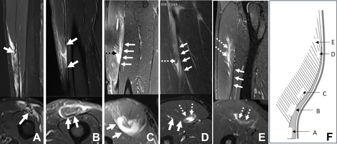
Figure 8.
Five different cases of posterior aponeurosis injury. Sagittal and axial T2-weighted fat-saturated magnetic resonance imaging scans of (A) a tendinous rupture (with posterior aponeurosis retracted), (B) a myotendinous rupture, (C) an intramuscular rupture, (D) a myoaponeurotic rupture, and (E) a myofascial injury. (F) Scheme in relation to these injuries. The white solid arrows indicate the posterior aponeurosis; the white and black dashed arrows indicate the injury.

