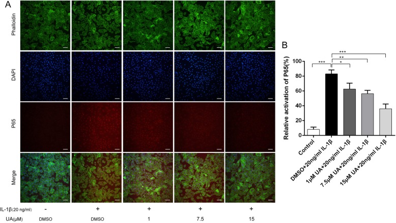
Fig. 5.
Effect of UA on IL-1β-induced nuclear translocation of P65. Chondrocytes were pretreated with UA (1, 7.5, and 15 μM) for 2 h, followed by co-incubation with 20 ng/ml IL-1β for 30 min and nuclear translocation of NF-κB P65 was detected by immunofluorescence. a The nuclear translocation of P65 detected by immunofluorescence (red signal represents P65, original magnification 100×, scale bar: 100 μm). b Relative activation of P65 shown as a histogram. Data are presented as mean ± S.D. n = 6. *P < 0.05, **P < 0.01, ***P < 0.001 versus the IL-1β group

