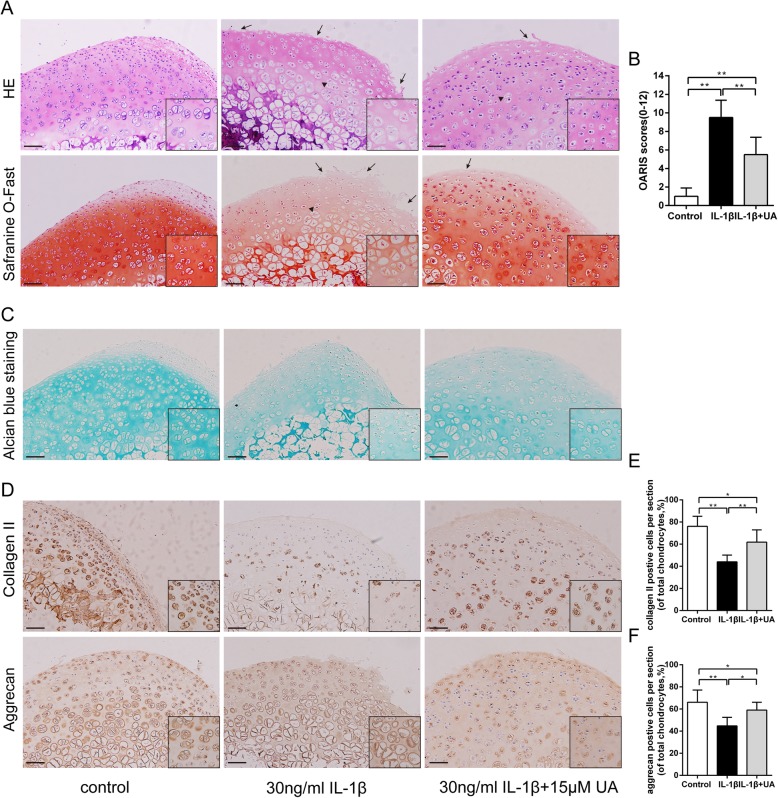
Fig. 6.
Effect of UA on IL-1β-induced cartilage damage in cartilage explants culture. Rat cartilage explants were exposed to 30 ng/ml IL-1β alone or with 15 μM UA for 3 days. a. Representative H&E and S-O Fast Green staining revealed the IL-1β treated group had apparent morphological changes including rough surfaces (black arrow), clustered and disorganized chondrocytes (black triangle), obvious hypocellularity, and loss of Safranin-O staining compared with the control group (scale bar: 20 μm). b. Diagram showing the OARSI scores of the cartilage. c. Alcian Blue staining for glycosaminoglycan (GAG) distribution (scale bar: 20 μm). d. Immunohistochemical staining of Collagen II, Aggrecan expression in the cartilage samples (scale bar: 20 μm). e and f. The percentages of Collagen II and Aggrecan positive cells in each section were quantified by Image Pro Plus. Three sections were randomly selected for quantification, and original magnification were 40 × and 200× in overall and partial picture, respectively. Data are presented as mean ± S.D. n = 6. *P < 0.05, **P < 0.01 versus the IL-1β group

