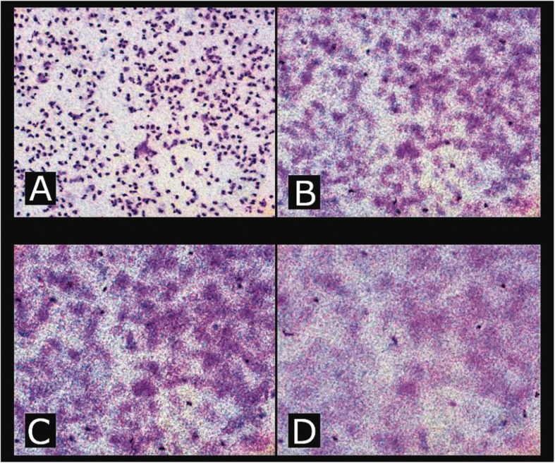
Fig. 1.

Leukocyte migration was evaluated using a Millipore membrane (Millipore). Migration was measured using a microscope where the Vernier graduated in micrometres (μm) was calibrated to obtain the measurements. The microscope was focused onto the cells on the upper surface of the membrane (a) and the position of the fine focus Vernier recorded. The depth of focus was then advanced through the membrane (b and c) until the last and furthest migrating cells were seen. At that position, the mark on the calibrated Vernier was again recorded (d). All photographs taken at 100× magnification
