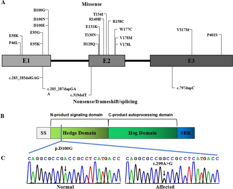
Fig. 4.
IHH pathogenic variants. a Boxes represent three different exons as indicated, and solid lines connecting these boxes represent the introns of IHH gene. The numbers above the boxes indicate the positions of the IHH complementary DNA at the start–stop sites and exon–intron boundaries. Vertical lines represent the locations of missense (above the boxes) or nonsense/frameshift/splicing (below the boxes) variants. b IHH protein structure with key domains, regions, and the mutation indicated. c Sanger sequencing chromatograms showing a missense variant c.299A > G(p.Asp100Gly) in the affected individuals in comparison to those of unaffected individuals

