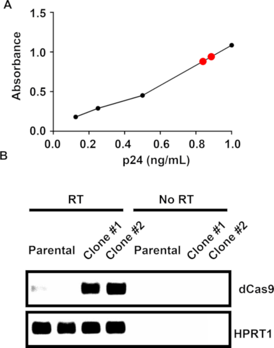
Figure 2: Viral titering and colony screening.
(A) An example p24 ELISA standard curve for lentivirus-containing, conditioned media. The black dots represent standard samples and the red dots unknown samples. (B) After transducing, selecting, and generating colonies, real-time PCR and gel electrophoresis against dCas9 were performed on the RNA from either nontransduced Jurkat cells (parental) or dCas9-transduced clones. No reverse transcriptase (No RT) controls were performed.

