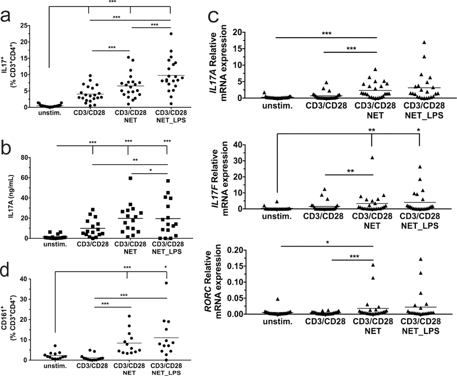
Figure 1. NETs promote CD3/CD28 induction of Th17 cells from PBMCs.
Unstimulated PBMCs, and PBMCs activated with anti-CD3/CD28 beads in presence of NETs or LPS-induced NETs, were incubated for 7 days followed by (a) flow cytometry to determine the percentage of IL-17+T-cells; (b) ELISA to determine secreted IL-17A protein; (c) qPCR to determine IL-17A, IL-17F and RORC mRNA levels; and (d) flow cytometry to determine the percentage of CD161+T-cells. Means are indicated by horizontal lines. Statistics: one-way repeated measures ANOVA was performed on transformed data (A and D, logit; B log (ln); C log2), with all p-values corrected to maintain experimentwise type I error rate for the tested factor. * indicates p < 0.05, ** indicates p < 0.01, *** indicates p < 0.001. Graphical results for the transformed data and a summary of statistically-significant outcomes are presented in Figure S1.

