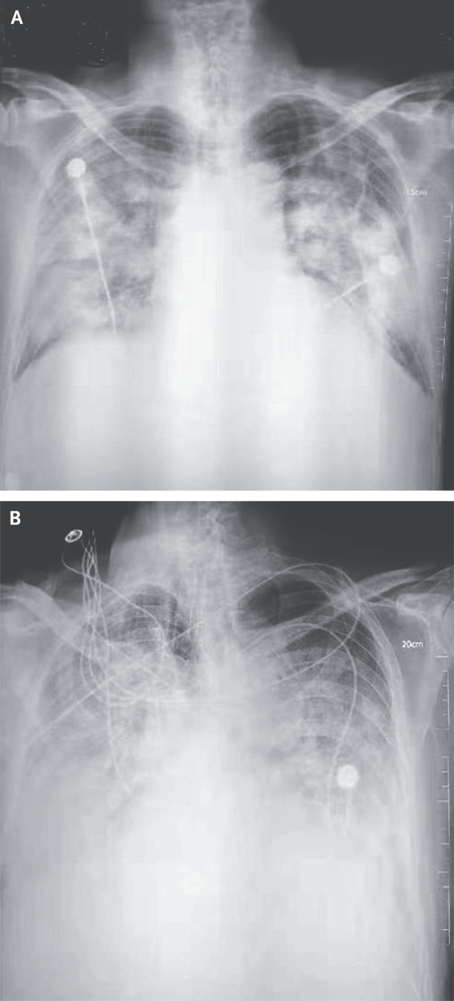
Figure 1. Chest Radiographs.
Shown are chest radiographs from Patient 2 on days 8 and 11 after the onset of illness. The trachea was intubated and mechanical ventilation instituted in the period between the acquisition of the two images. Bilateral fluffy opacities are present in both images but are increased in density, profusion, and confluence in the second image; these changes are most marked in the lower lung fields. Changes consistent with the accumulation of pleural liquid are also visible in the second image.

