
Fig. 3.
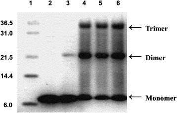
Chemical crosslinking of 2-helix protein. Cross-linked products were separated on Tris–Tricine SDS–PAGE followed by Coomassie brilliant blue staining. Protein markers (kDa) are shown in lane 1. Lanes 2–6 represent 0, 0.01, 0.1, 0.5, and 1.0 mM concentration of ethylene glycol bis (succinimidyl succinate) (EGS) used, respectively. Arrows corresponding to monomer, dimer, and trimer are indicated. They demonstrate that the 2-helix protein existed as trimer.