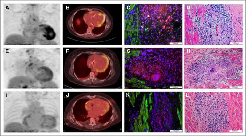
Figure. Cardiac positron emission tomography with 18F-fluorodeoxyglucose (FDG-PET) of patient No. 1 performed 1 mo before total artificial heart (TAH) showed severe-intensity FDG uptake in the apical septum and inferior walls.
A and B, An intense, positive staining for the ASC (apoptosis-associated speck-like protein containing a caspase recruitment domain) is shown in cardiac pathology tissue from the TAH surgery, which identifies the inflammasome (C). Classic hematoxylin and eosin (H&E) staining shows granulomas corresponding to inflammasome presence (D). FDG-PET of patient No. 2 performed 2 mo before left ventricular assist device (LVAD) showed moderate-intensity diffuse FDG uptake extending into the left ventricular apex (E and F). A moderate positivity for ASC in tissue from the LVAD apical core is displayed in G, and H&E staining shows a granuloma corresponding to inflammasome presence (H). The FDG-PET of patient No. 3 performed 1 mo before LVAD and 7 mo before orthotopic heart transplant showed mild-intensity patchy hypermetabolic activity in the left ventricle with mild activity in the distal lateral wall extending into the distal anterolateral wall and apex (I and J). A mildly positive staining for ASC of native cardiac tissue obtained during the transplant surgery is shown in K, and the corresponding H&E stain is shown in L.

