
Fig. 1.
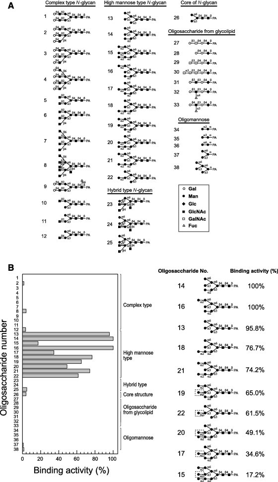
Oligosaccharide-binding specificity of KAA-2. (A) The structures of the pyridylaminated (PA)-oligosaccharides used in this study. Open circles, galactose; closed circles, mannose; closed diamonds, glucose; closed squares, N-acetylglucosamine; open squares, N-acetylgalactosamine; open triangles, fucose; open pentagons, xylose. (B) Binding activity of KAA-2 to PA-oligosaccharides. Binding activity was determined using the centrifugal ultrafiltration–HPLC method, and is expressed as the ratio (%) of the amount of bound oligosaccharide to that of the added oligosaccharide. The structures of the oligosaccharides that are selectively recognized by KAA-2 are shown. The non-reducing terminal α1–2 Man in the D2 arm (surrounded by a dotted line in the figure) negatively participates in binding to KAA-2. The assay was performed in duplicate for each PA-oligosaccharide and binding activity is expressed as the average value of duplicate assays.