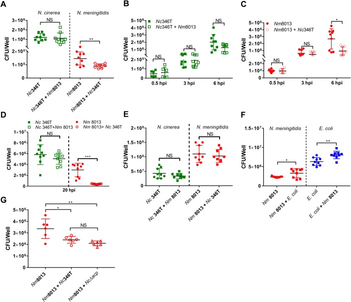
Fig 7. N. cinerea reduces association of N. meningitidis with epithelial cells.
(A) Cells were infected with N. cinerea (Nc346T) for 4.5 h prior to infection with N. meningitidis (Nm8013). The number of cell associated bacteria of each species was determined 1.5 h later. Results are the mean ± SD of three independent experiments carried out in triplicate. NS, not significant; ***p<0.0005 (unpaired two-tailed Student’s t-test). (B and C) Epithelial cells were infected with N. meningitidis (Nm 8013) alone or with N. cinerea (Nc 346T). The number of cell associated bacteria (CFU/mL) was determined at time points as indicated. Filled shapes show the number of CFU/well in single infections, while empty shapes show the number of CFU/well in co-infections. Each data point represents a single well from three independent experiments conducted in triplicate. NS, not significant; *, p<0.05 (unpaired two-tailed Student’s t-test). (D) Epithelial cells were infected with N. meningitidis (Nm 8013) alone or co-infected with N. cinerea (Nc 346T) at a ratio of 1:100 (Nm 8013 to Nc 346T) for 20 h. (E) Single and mixed cultures of N. meningitidis (8013) and N. cinerea (346T) were grown in the absence of cells for 6 hrs, and the number of bacteria was determined by selective plating. Results are the mean +SD of three independent experiments carried out in triplicate. NS, not significant. (F) Epithelial cells were infected with N. meningitidis (Nm 8013) alone or co-infected with E. coli (BL21 pET21b) at an MOI of 50 for each strain. Cell associated N. meningitidis and E. coli (CFU/well) was determined at 6 hpi. Filled circles show Nm8013 (red) and E. coli (blue) in single infections; filled squares show Nm8013 (red) and E. coli (blue) in co-infection. Results are the mean ± SD of 9 replicates from three independent experiments. NS, not significant; *p<0.05; **p<0.005 (unpaired two-tailed Student’s t-test). (G) Epithelial cells were infected with N. meningitidis (Nm 8013) alone or co-infected with wild-type N. cinerea (Nc346T) or a mutant lacking ACP (NcΔacp). At 6 hpi, cell associated N. meningitidis was quantified and presented as CFU per well. Filled circles show Nm8013 bacterial numbers in single infections; empty circles or empty triangles show levels of Nm8013 in co-infection. Results are the mean ± SD of at least three independent experiments carried out in duplicate. NS, not significant; *, p<0.01; **, p<0.001 (one-way ANOVA test for multiple comparison).

