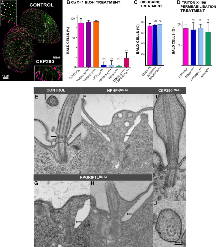
Fig 4. Depletion of CEP290, NPHP4, or RPGRIP1L affects deciliation ability and ciliary shape.
(A–D) Depletion of CEP290, NPHP4, or RPGRIP1L affects deciliation ability. (A) Control and CEP290-depleted cells were simultaneously submitted to deciliation treatment (5% EtOH, 1 mM Ca2+) prior to labelling cilia (TAP952 in magenta, polyE in green). Control cells were previously marked by India ink ingestion allowing their identification. Cep290RNAi cells bear cilia, whereas control cells are completely bald. Bar = 10 μm. (B) Bar plot showing the mean percentages of bald cells (less than 25% of cilia) after deciliation for control (n = 421 cells), TMEM107RNAi (n = 290 cells), TMEM216RNAi (n = 258 cells), CEP290RNAi (n = 98 cells), RPGRIP1LRNAi (n = 302 cells), NPHP4RNAi (n = 82 cells), and the double TMEM107/RPGRIP1LRNAi (n = 141 cells; error bars represent the standard deviation); >3 independent replicates per condition. Statistical significance was assessed by unpaired χ2 test. ****Two-sided p < 0.0001. Source data can be found in S3 Data. (C) Bar plot showing the mean percentages of bald cells (less than 25% of cilia) for control (n = 53 cells), CEP290RNAi (n = 47 cells), and RPGRIP1LRNAi (n = 68 cells) after deciliation with 5 mM dibucaine. Error bars represent the standard deviation. Three replicates per condition. Statistical significance was assessed by unpaired χ2 test two-sided p-value. Source data can be found in S3 Data. (D) Bar plot showing the mean percentages of bald cells (less than 25% of cilia) for control (n = 222 cells), CEP290RNAi (n = 77 cells), RPGRIP1LRNAi (n = 77 cells), and NPHP4RNAi (n = 76 cells) after detergent extraction using 0.01% Triton X-100. Note that in these conditions, more than 80% of cells deciliate. Three replicates per condition. Source data can be found in S3 Data. (E–J) Depletion of CEP290, NPHP4, and RPGRIP1L affects ciliary shape. EM images of abnormal cilia found in NPHP4RNAi (F), RPGRIP1LRNAi (G, H), and CEP290RNAi (I, J). A control cilium is shown in longitudinal and transverse sections in panel A. In NPHP4- and RPGRIP1L-depleted cells, the link between the axoneme and the ciliary membrane is missing at the level of the TZ. Abnormal cilia present extension defects, and vesicles accumulate inside the cilia, probably resulting from a defective TZ gate function. In CEP290-depleted cells, abnormal cilia present a strikingly enlarged lumen, resulting most probably from altered ciliary gate function. Bar: 200 nm. CEP290, centrosomal protein of 290 kDa; EM, electron microscopy; NPHP4, Nephronophtysis 4; polyE, anti-poly-glutamylated tubulin; RNAi, RNA interference; RPGRIP1L, Retinitis pigmentosa GTPase regulator-Interacting Protein 1-Like Protein; TMEM107, Transmembrane protein 107; TZ, transition zone.

