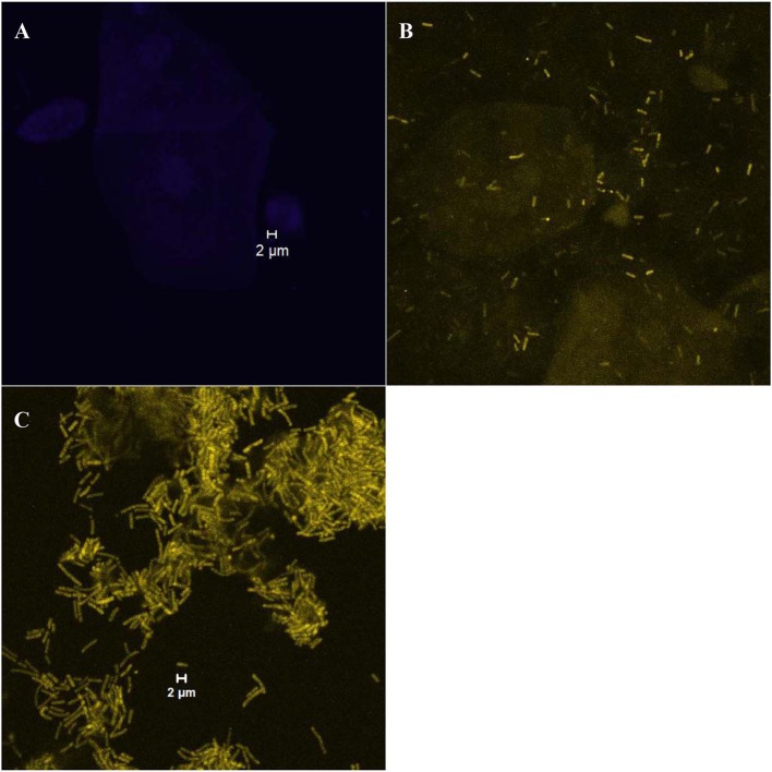
Figure 1.
Examples of different bacterial growth forms; (A) “absent” category with no bacteria present on vaginal cells (visualized with the laser diode 405 that detects the DAPI stain); (B) “dispersed” category [an example of scattered cells of Lactobacillus spp. (rods in yellow color) visualized with the helium-neon laser 2 that detects the Lab158-Cy3 probe]; (C) “biofilm” category ([an example of the Lactobacillus biofilm (rods in yellow color) visualized with the helium-neon laser 2]).

