
Fig. 3. Viremia of pregnant rhesus macaques after ZIKV inoculation.
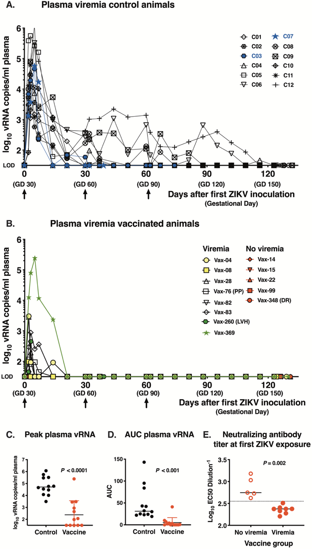
(A) Plasma ZIKV RNA (vRNA) in unimmunized control animals (n=12). Arrows show 3 ZIKV inoculations and blue shows the 2 animals with early fetal death. Each viremia value reported represents the mean of triplicate qRT-PCR measurements; the assay limit of detection (LOD) of ~1.5 log10 vRNA copies/ml is used as baseline for the Y-axis. (B) Plasma vRNA in vaccinated pregnant macaques (n=13). Yellow (n=2) and green (n=2) symbols indicate viremic animals for which the first ZIKV-inoculation occurred either very early (4–8 days) or late (260–369 days) after the 2nd DNA immunization, respectively. Red symbols are 5 vaccinated dams that after challenge had undetectable viremia. The three vaccinated animals with late fetal death are indicated: PP= placenta previa; LVH= left ventricular hypertrophy, DR=decidual reaction. (C) and (D). Peak viremia and area under the curve of log10-transformed plasma ZIKV RNA levels over time, respectively (both comparisons with two-tailed Mann Whitney test). (E) Neutralizing antibody titers (with indication of median) on day of first ZIKV challenge and viremia status of vaccinated pregnant animals (n=13) after ZIKV inoculation. The dotted line, drawn between the ‘not viremic’ and ‘viremic’ animals that were vaccinated is at EC50 dilution value of ~ 2.55 log10 (or titer of 1:355).