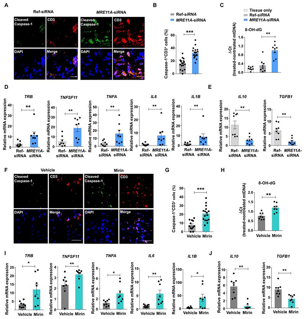
Figure 6: MRE11A regulates caspase-1 activation and mtDNA leakage in tissue-residing T cells.

NSG mice engrafted with human synovial tissue were reconstituted with healthy PBMCs. MRE11A was silenced in PBMCs before transfer or by daily injection of the MRE11A inhibitor Mirin (or vehicle). Control mice had human synovial tissue only. (A) Dual-color immunohistochemistry for cleaved caspase-1 (green) and CD3 (red) in synovial explants. Scale bar; 20 μM. (B) Frequencies of CD3+ T cells with cleaved caspase-1+ quantified in 18 random HPF. (C) Tissue mtDNA was extracted and 8-OH-dG was determined as in Figure 2F. (D, E) Tissue transcriptome analysis for inflammation-related genes by RT-PCR. (F) Representative microphotographs from tissue IHC stained for cleaved caspase-1 (green) and CD3 (red). Scale bar; 20 μM. (G) Percentages of CD3+ T cells expressing cleaved caspase-1 (12 random HPF). (H) 8-OH-dG measured in mtDNA extracted from synovial grafts. (I, J) Tissue transcriptome analysis for inflammation-related genes by RT-PCR. Mean ± SEM compared by Wilcoxon test. *p<0.05, **p<0.01, ***p<0.001. See also Fig.S6.
