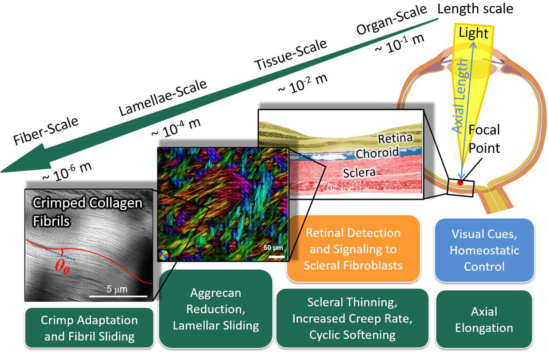
Figure 1:

Multi-scale mechanisms of scleral remodeling in myopia. The green boxes represent structural and material alterations due to remodeling; the blue box represents the primary stimulus; and the orange box represents stimulus detection and signaling pathways. Scleral remodeling during eye development is driven by visual cues and a homeostatic control mechanism that matches the eye’s axial length to its focal length at the organ-scale. Visual cues are detected by the retina, which sends signals through the choroid to the sclera to alter the scleral remodeling rate. Scleral remodeling in myopia leads to axial elongation at the organ-scale, scleral thinning, increased creep rate and cyclic softening at the tissue-scale. We propose that scleral remodeling involves lamellar sliding at the lamellae-scale, and fibril sliding and adaptation of the collagen fibril crimp at the fiber-scale. The colors at the lamellae-scale indicate the local fiber orientation, whereas the intensity is proportional to the collagen fiber density.
