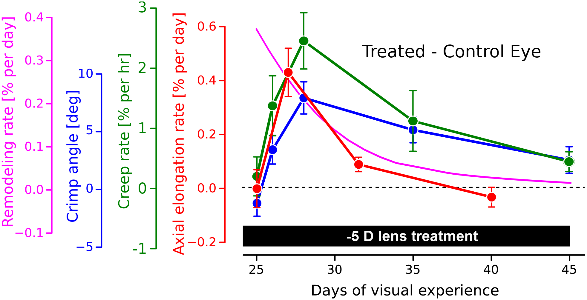
Figure 2:

Time-dependent changes in scleral biomechanics during experimentally induced myopia using a −5D lens in tree shrews. Plotted are the differences between lens treated and control eyes, showing a similar time-dependent trend for all of the study variables. The axial elongation rate (red curve, [20]) increases rapidly after lens placement, followed by a decline to normal levels as the eye adapted its axial length to the new focal length. The creep rate (green curve:, [20]) and the collagen fiber crimp angle (blue curve, [13]) show a similar time-dependent trend compared to the axial elongation rate. Based on the model assumptions, the computationally predicted remodeling rate (magenta curve, [22]) predicts an immediate increase in the remodeling rate after lens placement, followed by a gradual decrease that is consistent with the time-dependent biomechanical changes seen in the other curves.
