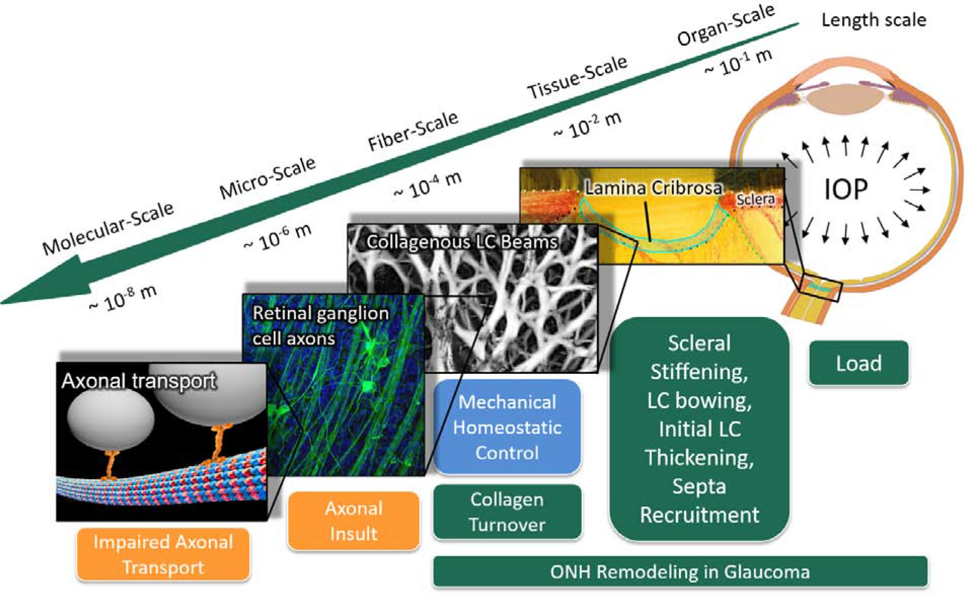
Figure 3:

Multi-scale mechanisms of ONH remodeling in glaucoma. The green boxes represent structural and material alterations due to ONH remodeling; the blue box represents the primary stimulus; and orange boxes represent mechanisms related to RGC axonal injury. The eye is subjected to IOP load at the organ-scale and chronic IOP elevation is the main risk factor for glaucoma. Pathologic ONH remodeling in glaucoma includes changes in scleral stiffness, LC bowing, initial LC thickening, and septa recruitment at the tissue-scale [31, 32, 33, 34, 35, 36]; and the synthesis of new LC beams at the fiber-scale [37]. We propose that these remodeling mechanisms are driven by a mechanical homeostatic control mechanism in an effort to maintain a homeostatic strain condition at the fiber-scale by altering collagen turnover in the ONH connective tissue. Pathologic ONH remodeling is thought to impair axonal transport by a direct or indirect mechanical insult to the RGC axons that pass through the LC porous microstructure. Fiber scale image is modified from a study by Brazile et al. [38]. ONH, optic nerve head; LC, lamina cribrosa; RGC, retinal ganglion cell; IOP, intraocular pressure.
