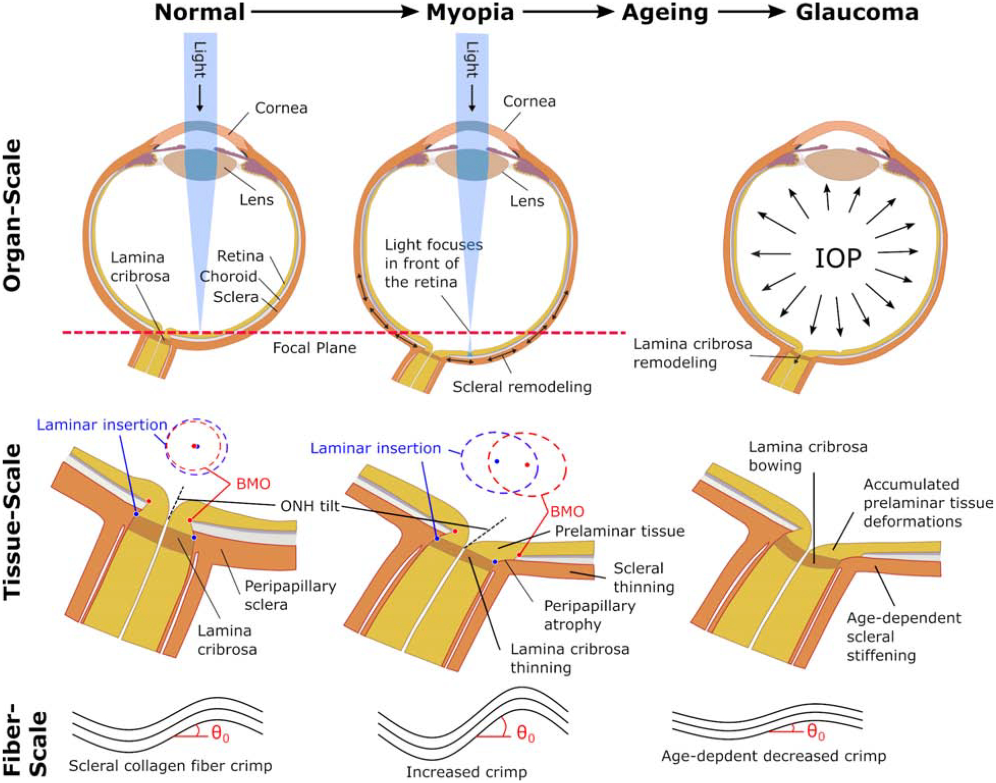
Figure 5:

Possible interactions at multiple scales between scleral and ONH remodeling in myopia, aging, and glaucoma. At the organ-scale, visual cues drive scleral remodeling that leads to axial elongation and myopia whereas IOP is thought to be the primary load that drives glaucomatous ONH remodeling in adults. At the tissue-scale, the vision-guided remodeling leads to asymmetric morphological changes of the ONH due to differential remodeling in the nasal/temporal sides. The LC and sclera thin as the LC shifts nasally with respect to the Bruch’s membrane opening (BMO). The blue and red ellipses represent en face views of the anterior laminar insertion and BMO, respectively, illustrating the increase in canal area, development of an elliptical canal shape, and relative deformations between the LC and BMO. These relative deformations lead to the tilted and rotated appearance of the myopic ONH, and potentially contribute to the development of peripapillary atrophy. Both, the sclera and the LC stiffen with age [46, 47]. In glaucoma, pathological ONH remodeling involves an initial thickening of the LC and posterior bowing of LC and sclera. ONH remodeling in myopia, age-dependent scleral stiffening, and pathologic remodeling in glaucoma, are all thought to promote glaucomatous LC bowing and tissue deformations in the prelaminar tissues. These prelaminar tissue deformations may accumulate as the eye develops myopia during childhood, ages, and develops glaucoma later in life. At the fiber-scale, the scleral collagen fiber crimp is temporarily increased during myopia development followed by a decrease with age. The age-dependent decrease in crimp angle θ0, is partially responsible for the increased scleral stiffness with age [45]. Experimental evidence supports the notion that mechanical homeostatic conditions are defined at the fiber-scale. ONH, optic nerve head; IOP, intraocular pressure; LC, lamina cribrosa.
