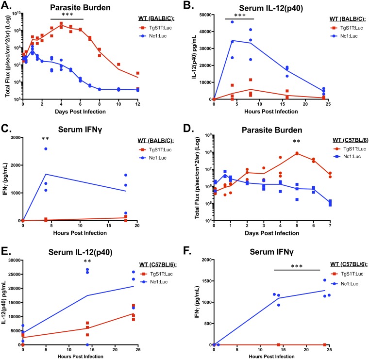
FIG 2.
Parasite burden and day 0 to 1 cytokine levels in mice infected with N. caninum (Nc1) or T. gondii (S1T). Six-week-old BALB/c (A to C) or C57BL/6 (D to F) mice were i.p. injected with 106 TgS1T:Luc or Nc1:Luc tachyzoites. Bioluminescence imaging was used to monitor the parasite burden over the course of the infection. Serum was collected and analyzed for IL-12p40 or IFN-γ by an ELISA. (A) Quantification of bioluminescence during in vivo infections in BALB/c mice (TgS1T:Luc, n = 2; Nc1:Luc, n = 3) (the experiment was repeated [see Fig. S1 in the supplemental material]). (B and C) Cytokine quantification using an ELISA for mouse IL-12p40 (B) or IFN-γ (C) (n = 3 per parasite species). (D to F) Same as panels A to C but with C57BL/6 mice (n = 3 per species; this experiment was performed only once). For all experiments, imaging data were log transformed, and two-way repeated-measures ANOVA (alpha value of 0.05) with Sidak’s multiple-comparison test was then performed. Cytokines were analyzed by two-way repeated-measures ANOVA (alpha value of 0.05) with Sidak’s multiple-comparison test. **, P < 0.01; ***, P < 0.001.

