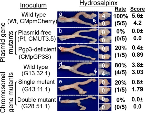
FIG 2.

Detecting gross pathology from mouse genital tract following intravaginal inoculation with chlamydial organisms with or without mutations. Groups of CBA/J mice (n = 5) intravaginally inoculated with different C. muridarum organisms as described in the Fig. 1 legend (panel a for mice infected with CMpmCherry, panel b for Pf, CMUT3.5, panel c for CMpGP3S, panel d for C. muridarum wild-type clone G13.32.1, panel e for mutant clone G13.11.1, and panel f for mutant clone 28.51.1). On day 56 after infection, all mice were sacrificed for observing hydrosalpinx pathology, as described in Materials and Methods. One representative genital tract image from each group is shown with the vagina on the left and oviduct/ovary on the right. Oviducts with hydrosalpinges are indicated by white arrows. The oviduct/ovary from each side is magnified in the panels on the right, with the corresponding hydrosalpinx scores marked. The hydrosalpinx was counted and semiquantitatively graded to calculate the incidence rate and severity score in a given group.
