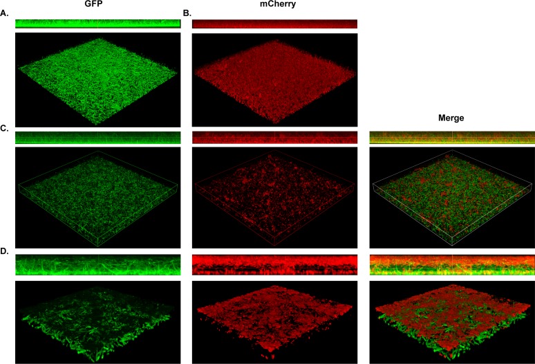
FIG 3.
S. maltophilia and P. aeruginosa stratify within polymicrobial biofilms in vitro. Structural composition of polymicrobial biofilms was assessed via confocal imaging of S. maltophilia K279a (GFP+), P. aeruginosa mPA0831 (mCherry+), or both grown at 30°C for 8 hours. Single-species biofilms of S. maltophilia K279a (GFP+) (A) and P. aeruginosa mPA0831 (mCherry+) (B) were imaged at ×40 magnification. Dual-species foci were imaged at ×40 magnification (C) and zoomed 4× for a total magnification of ×160 (D).

