FIG 5.
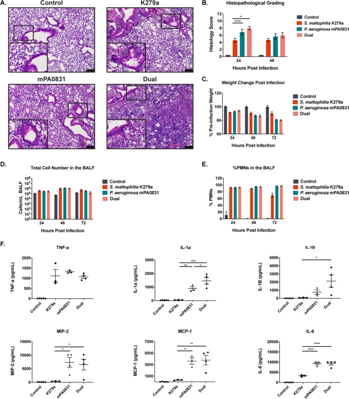
Inflammatory consequences of polymicrobial infection closely resemble those of P. aeruginosa single-species infection. (A) Representative images were taken of H&E-stained lung sections from mice infected with S. maltophilia K279a and P. aeruginosa mPA0831 alone and in combination. (B) Severity of infection as indicated by H&E staining was graded via a semiquantitative histology score for 24 and 48 hours postinfection. Mean ± SEM, n = 3 to 8. (C) Change in weight following infection was monitored. Mean ± SEM, n = 4 to 6. (D and E) Total immune cell number (D) and percentage of PMNs in the BALF (E) were quantified via differential cell counts. Mean ± SEM, n = 3 to 5. Two-way ANOVA with Tukey’s post hoc comparisons; *, P < 0.05; ****, P < 0.0001. (F) Cytokine/chemokine analysis performed on BALF supernatant using a Luminex multiplex assay. Mean ± SEM, n = 3 to 4. One-way ANOVA with Tukey’s multiple-comparison test for post hoc analysis. *, P < 0.05; **, P < 0.01; ***, P < 0.001; ****, P < 0.0001. Significant outliers were identified via ROUT method and removed.

