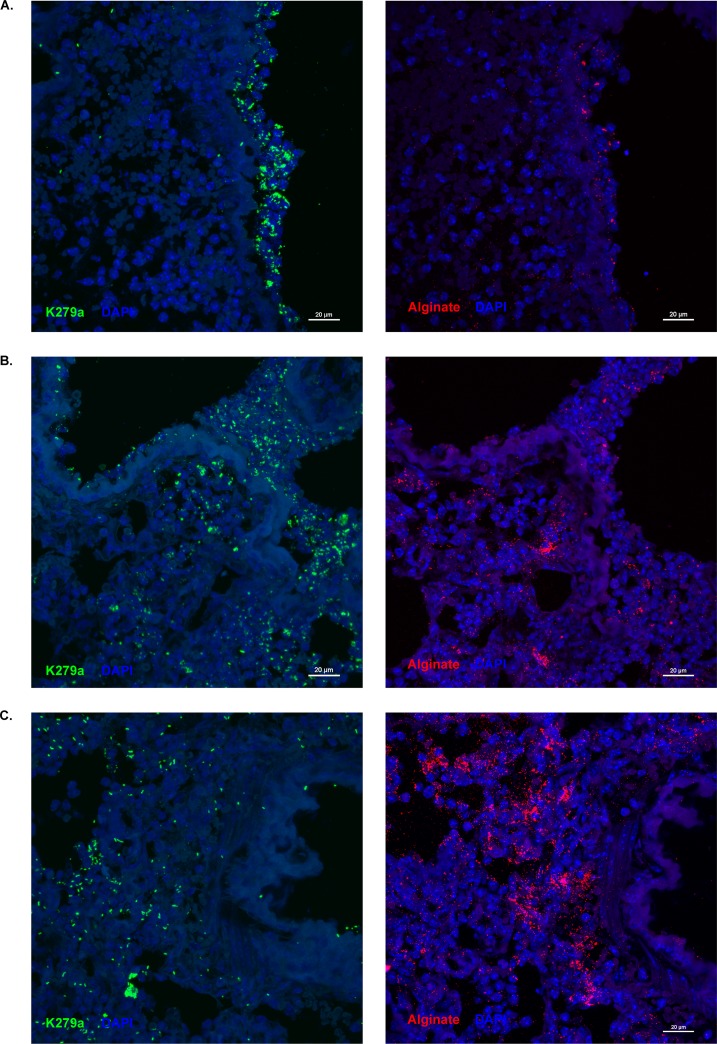
FIG 7.
S. maltophilia colocalizes with alginate during polymicrobial infection in the lung. Representative images of fluorescently stained lung sections were taken via confocal laser scanning microscopy (CLSM). Ten-micrometer serial sections were cut from lungs of mice dually infected with ∼107 CFU/mouse of S. maltophilia K279a and P. aeruginosa mPA0831. Lung sections from dual species-infected mice were stained for S. maltophilia via antisera from rabbits immunized with heat-killed S. maltophilia (green) and for alginate via an anti-alginate polyclonal rabbit antibody (red). Lung structures were visualized with 4′,6-diamidino-2-phenylindole (DAPI; blue). Dual-species foci were imaged on the airway surface (A) and in the lung parenchyma (B and C) at ×40 magnification.

