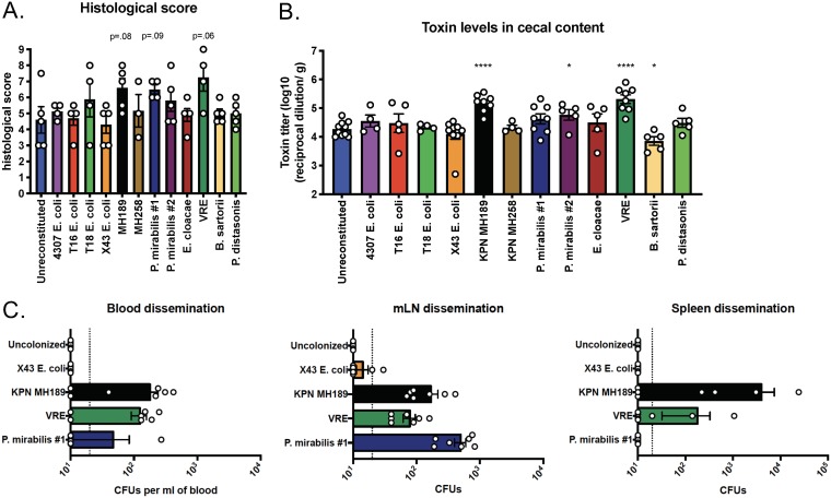
FIG 6.
Impact of the residual microbiota on host pathology, cecal toxin levels, and bacterial dissemination during CDI. The experimental design follows the schematic in Fig. 5A. (A) Pathology scores of histological tissue sections based on cellular infiltration, edema (host response), and epithelial layer damage. Data shown are means ± SEM. (B) C. difficile toxin titers in cecal contents were determined by an in vitro cytotoxicity assay. *, P < 0.05; ***, P < 0.001; ****, P < 0.0001. (C) Mice (n, 7 from two independent experiments) were colonized with one of four strains. The mice were inoculated with 200 spores of C. difficile VPI 10463 and were euthanized on day 2 post-C. difficile infection. Blood, mesenteric lymph nodes (mLNs), and spleens were isolated and homogenized, and bacterial dissemination post-C. difficile infection was determined via selective plating.

