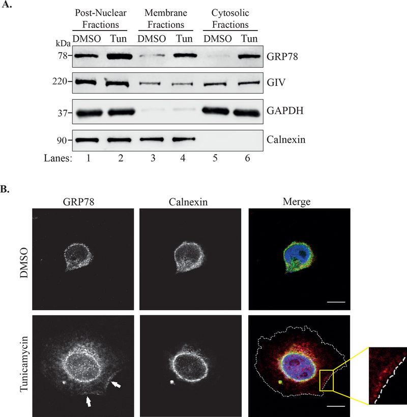
Figure 2. GRP78 localization is not restricted to the ER during ER stress.
A. Lysates of HeLa cells treated with either DMSO or tunicamycin for 6h were separated into cytosolic and membrane fractions by differential centrifugation. The fractions were analyzed by immunoblotting. GAPDH is used as a cytosolic marker and Calnexin as an ER marker. B. HeLa cells treated with tunicamycin or DMSO for 6h were fixed and stained for GRP78 (red), calnexin (green) and DAPI (blue). Cells were visualized by confocal microscopy. The arrows point to the localization of GRP78 along the cell periphery. The cell periphery is outlined by the dashed white line in the bottom, merged panel. The inset shows enlargement of the boxed region. Scale bar = 10 μm.

