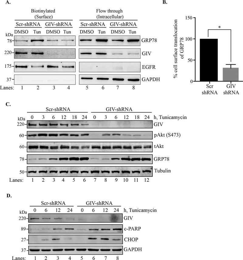
Figure 4. ER stress induced cell surface localization of GRP78 and Akt signaling is reduced in GIV depleted cells.
A. Scr-shRNA and GIV-shRNA cells were treated with tunicamycin or DMSO for 6h following which the cell surface proteins were biotinylated and separated from the non-biotinylated fraction through affinity purification using the streptavidin-agarose resin. The biotinylated (cell surface proteins) and flow-through (intracellular) fractions were then analyzed by immunoblotting. EGFR served as a cell surface protein control, and GAPDH as an intracellular protein control. B. The % cell surface localization of GRP78 was calculated by densitometric analysis. The Scr control was plotted to 100% and the relative % localization in GIV-shRNA cells was calculated. n=3. Errors bars represent ± S.E.M. *p <0.05. C. and D. Scr-shRNA and GIV-shRNA cells were ER stressed using tunicamycin for the indicated time-points before lysis. The cell lysates were analyzed by immunoblotting.

