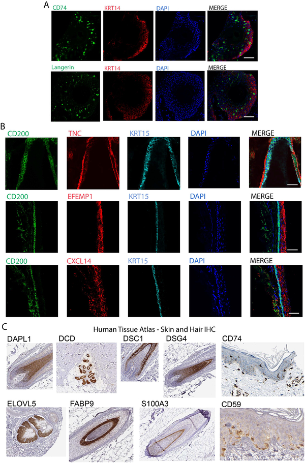
Figure 3. Validation of enrichment of gene expression at the protein level.
(A) Immunostaining of serial sections of frozen hair follicle samples labeled with CD74 and Langerin (green) highlight Langerhans cells in the basal interfollicular epidermis (KRT14+, red). Nuclei labeled with DAPI. (B) Immunostaining of serial sections of frozen hair follicle samples labeled with antibodies against CXCL14, TNC, EFEMP1 (red) compared to canonical bulge markers CD200 (green) and KRT15 (blue). Nuclei labeled with DAPI. Note that scale bars for (A) and (B) indicate 50uM. (C) Immunohistochemistry the indicated epitopes derived from the Human Tissue Atlas for markers identified by scRNA-seq as specific to particular cell types.

