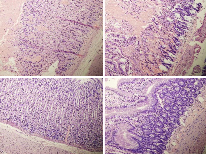
Figure 10.
Histological presentation in rats with suprahepatic ligation of the inferior vena cava. Assessment (stained with hematoxylin-eosin, magnification x 10, scare bar: 50 µm) was at 48 h post-ligation, in controls (upper) and BPC 157-treated rats (lower), stomach (left upper, control; lower, BPC 157), duodenum (right upper, control; lower, BPC 157). Note, substantial congestion and dilatation of mucosal and submucosal capillaries, submucosal edema, ischemic changes, such as architectural distortion and foci of hemorrhage with fibrin deposition in controls (upper), which were markedly counteracted in BPC 157-treated rats (lower).

