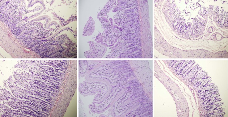
Figure 11.
Histological presentation in rats with suprahepatic ligation of the inferior vena cava. Assessment (stained with hematoxylin-eosin, magnification x 10, scare bar: 50 µm) waste 48 h post-ligation, in controls (upper) and BPC 157-treated rats (lower), jejunum (left upper, control; lower, BPC 157), ileum (middle upper, control; lower, BPC 157), cecum (right upper, control; lower, BPC 157). Note, substantial capillary congestion with mild ischemic changes, loss of crypts with foci of haemorrhage, edema of the lamina propria and mild lymphocytic infiltration in controls (upper), which were markedly counteracted in BPC 157-treated rats (lower).

