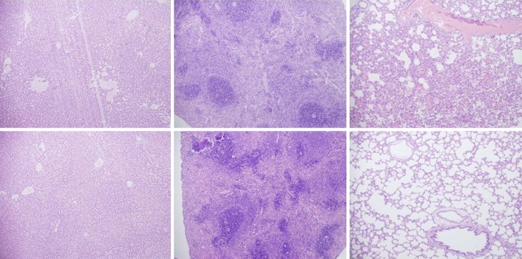
Figure 9.
Histological presentation in rats with suprahepatic ligation of the inferior vena cava. Assessment (stained with hematoxylin-eosin, magnification x 10, scare bar: 50 µm) was made at 48 h post-ligation, in controls (upper) and BPC 157-treated rats (lower), liver (left upper, control; lower, BPC 157), spleen (middle upper, control; lower, BPC 157), and lungs (right upper, control; lower, BPC 157). Note, the substantial congestion of the central vein, branches of the terminal portal venules, and sinusoidal dilatation in liver, sinusoidal congestion, dilatation and enlargement of red pulp leading to reduction of white pulp in spleen, edema of the interstitium, substantial dilatation and congestion of capillaries in the alveolar septum in lungs (upper), which were markedly counteracted in BPC 157-treated rats (lower).

