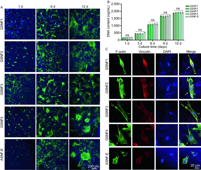
Figure 4.
Cytocompatibility and cell adhesion on the hydrogels with mechanical cues. (A) Confocal microscopy images of BMSCs on the hydrogels when cultured for 1, 6, and 12 days; (B) BMSC proliferation on different hydrogels when cultured for 1, 3, 6, 9 and 12 days; (C) immunofluorescence staining of adhered BMSCs on the hydrogels for 24 h. Nuclei is stained in blue. Vinculin is stained in red, and F-actin is stained in green. The white arrows indicate the oriented spreading of cells. ns means none statistically significant

