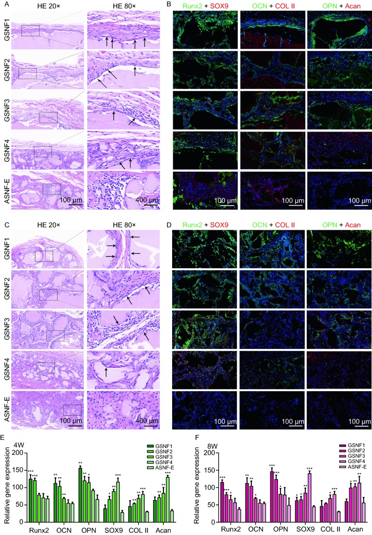
Figure 6.
Heterotopic ossification of hydrogels with gradient mechanical cuesin vivo. (A) H&E staining of the hydrogels when implanted subcutaneously for 4 weeks, the black arrows indicate the oriented growth of cells around the GSNF hydrogels; (B) Immunofluorescence analysis of osteogenic/chondrogenic-related markers of the hydrogels when implanted subcutaneously for 4 weeks. Cell nuclei are stained in blue, Runx2, OCN, and OPN are stained in green, SOX9, COL II and Acan are stained in red. Scale bar 100 μm; (C) H&E staining of the hydrogels when implanted subcutaneously for 8 weeks; (D) Immunofluorescence analysis of osteogenic/chondrogenic-related markers of the hydrogels when implanted subcutaneously for 8 weeks. Cell nuclei are stained in blue, Runx2, OCN, and OPN are stained in green, SOX9, COL II and Acan are stained in red. Scale bar 100 μm; (E and F) Quantitative analysis of fluorescence intensity of osteogenic/chondrogenic specific proteins when implantation for 4 and 8 weeks. *P ≤ 0.05, **P ≤ 0.01, and ***P ≤ 0.001

