Figure 2.
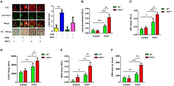
SHIP-1 deficiency aggravates lipid raft aggregation, oxidative damage, and bacterial dissemination after PAO1 infection. (A) MH-S cells were transfected with SHIP-1 siRNA (or negative control siRNA) for 48 h or treated with lipid raft blocker MβCD at 10 mM, then infected with green fluorescent protein (GFP)–PAO1 for 30 min, and stained with lipid raft marker (TRITC-labeled cholera toxin B, CTB-red) at 1/10,000 dilution; images were captured using microscope and assessed using ImageJ software on the basis of minimal 100 cells; arrows indicate the extent of lipid raft aggregation. (B) Peroxidation product was measured by a colorimetric assay in the lung tissue lysate. (C) Superoxide production in bronchoalveolar lavage fluid (BALF) cells significantly increased in PAO1-infected SHIP-1−/− mice compared with WT mice using the nitroblue tetrazolium (NBT) assay (1 mg/ml) at 560-nm absorbance. (D) Oxidative stress was increased in SHIP-1−/− AM cells as determined by the H2DCF fluorescence assay (5 μM) at 488 nm. (E) Myeloperoxidase (MPO) was measured from the lungs of SHIP−/− mice as compared with WT mice. (F) Polymorphonuclear neutrophils (PMNs) were significantly increased in infected SHIP-1−/− mice compared with WT mice (the data are mean ± SEM and are representative of three independent experiments, one-way ANOVA with Tukey post hoc test, *p < 0.05, **p < 0.01, ***p < 0.001).

