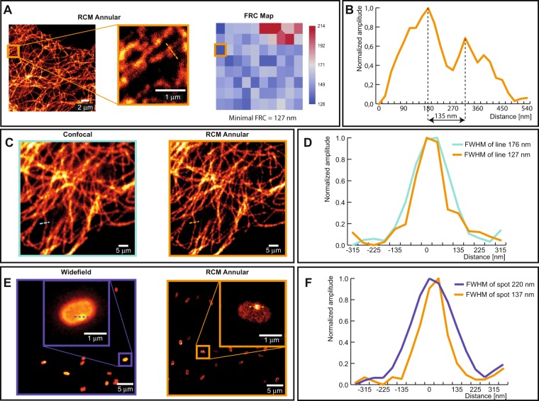
Figure 3.
Images of tubulin and a Bacillus subtilis spore using conventional approaches and RCM with annular illumination. (A) RCM Annular image of a tubulin along with a corresponding map of Fourier ring correlations (FRC) obtained by using the ImageJ NanoJ-SQUIRREL plugin. (B) Line profile plot indicating feature seperation of two tubulin filaments by a distance of 135 nm. (C) Comparison of a confocal image (left) and an RCM Annular image (right) of tubulin stained with Alexa 488 in HeLa cells. (D) Measured FWHM from the spatial intensity profile of a tubulin. (E) Comparison of a widefield Fluorescence image of an RCC PS832_GerKB-sGFP dormant spore (left) and RCM Annular microscopy (right). (F) GerKB-sGFP in a wild-type spores were visualized as a spot with a FWHM 137 nm by RCM Annular microscopy.

