Figure 1.
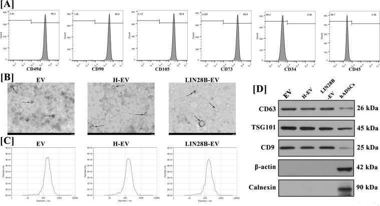
Characterisation of hADSCs and hADSC-EVs. (A) The expression of hADSCs surface markers CD49d, CD34, CD45, CD90, CD105 and CD73 was measured by flow cytometry. hADSCs were highly positive for CD105, CD90, CD73 and CD49d but negative for CD34 and CD45. (B) The size and the spheroid morphology of hADSC-EVs, hypoxic hADSC-EVs and LIN28B transfected hADSC-EVs (pointed by Black arrows) were shown under TEM (×25,000; 80 kV); scale bar: 500 nm. (C) The particle size of hADSC-EVs was assessed by NanoSight. NanoSight indicated that the average particle sizes of normal, hypoxic and LIN28B transfected hADSC-EVs were 183.1 ± 15.3 nm, 180.7 ± 14.9 nm and 174.5 ± 17.8 nm. (D) Western blot analysis indicated that hADSC-EVs had high expression of the surface markers of EVs, including CD63, CD9 and TSG101, which were relatively lower expressed in hADSCs. hADSC-EVs did not express β-actin and calnexin, which were higher expressed in hADSCs.

