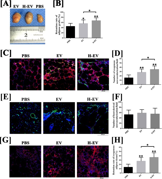
Figure 2.
Animal model of fat grafting (n = 12). (A) The fat grafts harvested at post-grafted week 12. (B) Line charts were plotted to compare the volume retention rate of fat grafts at post-grafted week 12. (C,D); The results of CD31(green)/human mitochondria immunofluorescence(red) staining indicated that, at week 4 post-transplantation, increased exogenous angiogenesis was observed in fat grafts co-transplanted with hADSC-EVs and hypoxic hADSC-EVs, compared with those co-transplanted with PBS. Moreover, co-transplanting with hypoxic hADSC-EVs can further enhance the exogenous angiogenesis in the early stage, compared with co-transplanting with normal hADSC-EVs. Scale bar: 200 μm. (E,F) The results of immunofluorescence staining (CD31/human mitochondria) indicated that host-derived CD31-positive vessels (human mitochondria-) were observed in the peripheral region of fat grafts. Semi-quantitative analysis for the calculation of host-derived neovascularisation indicated that co-transplantation with hADSC-EVs or hypoxic hADSC-EVs exerted no significant effect on host-derived angiogenesis at post-grafted week 4, compared with those co-transplanted with PBS. Scale bar: 200 μm. (G,H) The results of immunofluorescence staining (human mitochondria) indicated that, at week 12 post-transplantation, significant increases in retention rate of exogenous adipose tissue were observed in grafts co-transplanted with hADSC-EVs and hypoxic hADSC-EVs, compared with that co-transplanted with PBS. The retention rate of exogenous adipose tissue can be further improved by co-transplanted hypoxic hADSC-EVs compared with normal hADSC-EVs. Scale bar: 500 μm. Data are presented as the mean ± SD (n = 12). *P < 0.05, **P < 0.01.

