Figure 3.
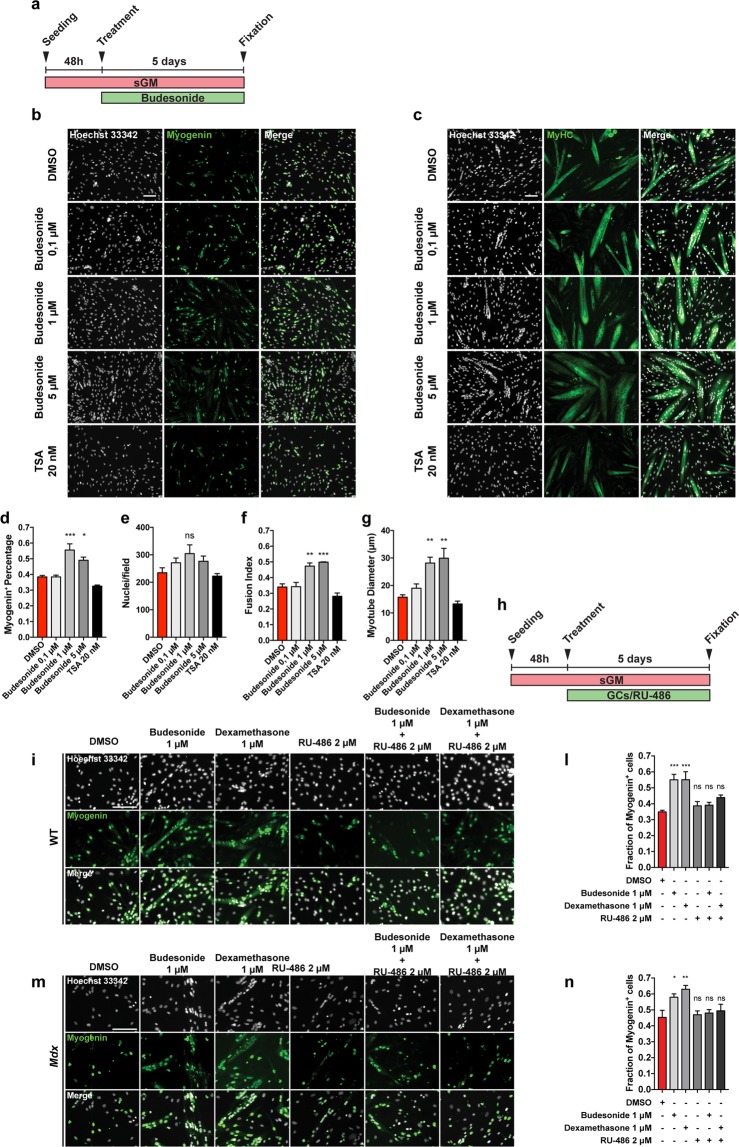
Budesonide treatment promotes terminal differentiation of mdx satellite cells through the activation of GCr. (a) SCs were isolated from muscles of mdx mice as CD45-/CD31-/ITGA7 + cells and plated in sGM. 48 hours after plating, cells were treated with three concentrations of budesonide (0.1, 1 and 5 μM) or TSA (20 nm) for 5 additional days. Myogenic differentiation was assessed by immunostaining with antibodies against myogenin (b) and MyHC (c) as late muscle-specific differentiation markers. Nuclei were counterstained with Hoechst 33342. (d) Column chart showing the percentages of myogenin positive cells in the experiment in panel b. (e) Bar plot reporting the number of nuclei per field for the experiments in b and c. (f,g) Bar plots showing the fusion index and myotube diameter for the experiment in c. The values are mean of three independent experiments ± SEM (n = 3). Statistical significance was evaluated using one-way ANOVA (*p ≤ 0.05, **p ≤ 0.01, ***P ≤ 0.001, ns: not significant). Scale bar: 100 μm. (h) Schematic representation of the experiments represented in i and m. 48 hours upon seeding, WT or mdx SCs were treated with budesonide, RU-486 or a combination of both. (i,m) Differentiating SCs were detected with an antibody against myogenin (green) and nuclei were counterstained with Hoechst 33342 (grey). (l,n) Bar plot showing the fraction of myogenin positive cells for the experiments reported in i and m. The values are mean of three independent experiments ± SEM (n = 3). Statistical significance was evaluated using one-way ANOVA (*p ≤ 0.05, **p ≤ 0.01, ***p ≤ 0.001, ns: not significant). Scale bar: 100 μm.

