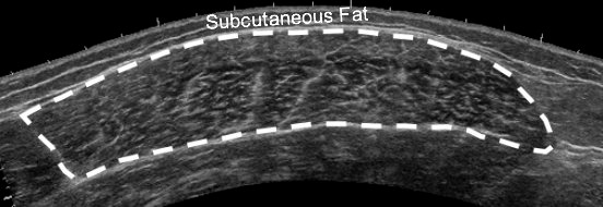
Figure 1.

Example of a panoramic ultrasound image of the quadriceps muscle. The interface between the hyperechoic epimysium and hypoechoic muscle tissue was traced, and the resulting area was defined as the vastus lateralis cross-sectional area. Echo intensity was derived from the average grayscale of all the pixels within the region of interest.
