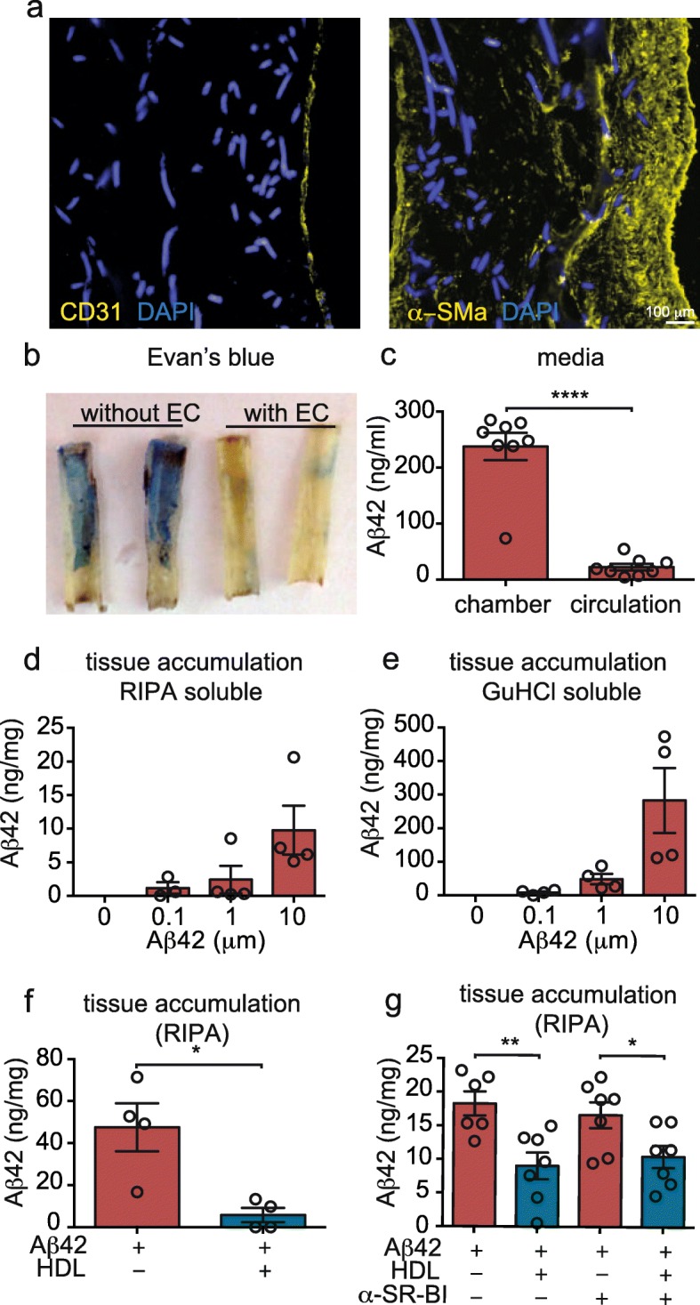
Fig. 1.

Human in vitro bioengineered vessels model CAA. a Immunofluorescent staining for CD31 confirms the formation of an endothelial monolayer surrounded by several layers of SMC stained for α-SM actin. b Evans blue staining confirms a tight endothelial barrier 10 days after endothelialisation. In (a) and (b), autofluorescence of the scaffold material is visible in the DAPI channel. c After injecting 1 μM of Aβ42 in the antelumen (tissue chamber), tissue chamber and circulation media were collected and Aβ42 was quantified by ELISA. d-e 24 h after injection of the indicated Aβ42 concentration into the tissue chamber, tissues were lysed in RIPA buffer before solubilizing the pellet in GuHCl followed by Aβ42 quantification using ELISA. f After injecting 1 μM of Aβ42 in the tissue chamber, 200 μg/mL of HDL was circulated through the lumen. After 24 h, Aβ42 accumulation was measured as above. g A blocking antibody against the HDL binding protein SR-BI was circulated in the presence of 200 μg/mL of HDL or BSA through the lumen after injecting Aβ42 into the tissue chamber. Aβ42 accumulation was measured as above. Points in graphed data represent individual bioengineered vessels, bars represent mean, error bars represent ±SEM and analysed by Student’ t-test or one way ANOVA *P < 0.05, **P < 0.01, ***P < 0.001 and ****P < 0.0001
