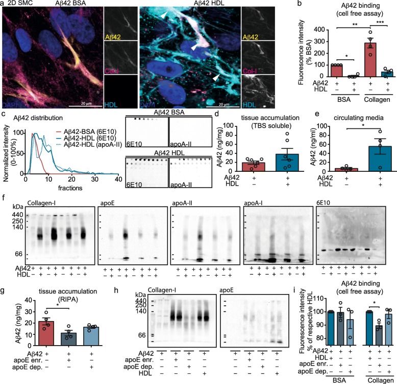
Fig. 5.
HDL binds to both collagen-I and to Aβ42 to form a complex and increases Aβ transport through the bioengineered vessel wall primarily by HDL-apoE particles. a SMC were grown for 3 d on 2D coverslips before incubating with 0.5 μM FITC-Aβ42 (depicted as yellow) and 200 μg/ml Alexa-633 labeled HDL (depicted as cyan). After 24 h, cells were fixed and stained for collagen-I (magenta) before imaging by confocal microscopy. White arrows show co-localization of Aβ42, HDL and collagen-I. b Black 96 well-plates were coated either with 50 μg/mL rat-tail collagen-I or 10% BSA as protein control for 24 h before incubating with a solution of 1 μM of FITC-Aβ42 with or without 200 μg/mL of HDL. After 24 h and extensive PBS washes, fluorescence representing bound Aβ42 was measured at 520 nm, excitation 490 nm. c 1 μM Aβ42 were incubated either with 200 μg/mL of HDL or BSA for 24 h at 37 °C before gel-filtration chromatography separation. Fractions were dot blotted and immunodetected for Aβ (6E10) or HDL (apoA-II). The intensity of each fraction on the dot blot was quantified, normalized between 0 and 100% and graphed on the left panel. The right panel shows a representative dot blot used for quantification. d 1 μM Aβ42 was injected in the tissue chamber of bioengineered vessels and 200 μg/mL of HDL was circulated through the lumen. After 24 h, tissues were homogenized in TBS to collect soluble Aβ, which was quantified in the tissue (c) or circulating media (d) by ELISA. f 1 μM Aβ42 was injected in the tissue chamber of bioengineered vessels and 200 μg/mL of HDL was circulated through the lumen. After 24 h vessels were lysed in RIPA and protein distribution was analysed using non-denaturing native blot probed for collagen-I, apoE, apoA-II, apoA-I and Aβ (6E10). g HDL was enriched for or depleted of apoE using an apoE immunoaffinity column. Bioengineered vessels were then treated with either fraction (200 μg/mL) in the presence of 1 μM Aβ42 as above before measuring Aβ deposition in the RIPA-soluble fraction. h RIPA lysates from engineered vessels were then analysed using non-denaturing native blot probed for collagen-I and apoE. i Black 96-well plates were coated either with 50 μg/mL rat-tail collagen-I or 10% BSA for 24 h before incubating with a solution of 1 μM of FITC-Aβ42 with or without 200 μg/ml of total HDL, apoE-depleted HDL or apoE-enriched HDL. After 24 h and extensive PBS washes, fluorescence was measured at 520 nm, excitation 490 nm. FPLC data and Western blots are representative of at least 3 individual experiments. Points in graphed data represent individual bioengineered vessels, bars represent mean, error bars represent ±SEM and analysed by Student’s t-test or one way ANOVA *P < 0.05, **P < 0.01, ***P < 0.001 and ****P < 0.0001

